

What types of brachytherapy can be used for treatment of this cancer type?
Brachytherapy delivers the radiation with a high degree of precision. This accuracy allows positioning and the calculation of the depth of irradiation so that healthy tissue is spared whilst the tumor gets a high dose of radiation.1 This increases efficacy and minimizes damage to healthy surrounding tissues.
Two types of brachytherapy are currently available for skin cancer:1
- Superficial, also called contact brachytherapy.
- Interstitial, with the insertion of plastic tubes or rigid needles.
Superficial brachytherapy involves moulds (often custom-made) and flaps for larger lesions, and radionuclide or electronic based shielded applicators for small volume lesions. Interstitial brachytherapy is applied to deeper located and/or very irregular shaped tumors.1

Brachytherapy not only provides good cosmetic results but has also been shown to be highly effective in preventing the skin cancer from returning.
When compared to external beam radiation therapy, with the source placed at a distance, brachytherapy reduces the amount of radiation to healthy tissues and can be delivered in a much shorter treatment time.1-3
Both basal cell and squamous cell cancers that have not spread to other parts of the body can be treated effectively with brachytherapy. Absolute contraindications for brachytherapy in skin cancer are invasion of bones, some genetic diseases like xeroderma pigmentosa, extension of the tumor in the orbit and perineural invasion.4

How brachytherapy for this cancer type is performed.
There are three main stages to the brachytherapy procedure: a) planning, b) treatment delivery and c) post-procedure monitoring.
Planning
The planning stage involves a thorough examination of the skin cancer and surrounding area. Ultrasound and/or biopsy may be used to gain an accurate picture of the layers of the skin and the precise position and thickness of the tumor.
The doctor calculates how much radiation is needed to treat the cancer and where the radiation should be placed over the skin.
In some cases, especially with larger lesions or lesions on a very irregular surface a mould of the skin may be taken. This enables the brachytherapy team to create a custom-made device to accurately place the radiation on the skin.
Treatment delivery
Applicator placement

Usually a patient is positioned on a bed or treatment chair. The doctor may use a special pillow to help the patient to maintain a stable position. Then the doctor will precisely position the treatment applicator on the area of skin to be treated.
The role of the skin applicator is to hold the source of radiation in the correct place over the skin during the ‘radiation delivery’ stage and to reduce the radiation to other nearby areas.
There are many different types of applicator available, and the exact type will depend on which type of brachytherapy is applied and on the size and shape of the lesion.
The applicators are designed to provide a close and reproducible fit to the surface of the skin.
In some cases a custom-made applicator will be used from a plaster mould of the skin.
Picture left: Electronic brachytherapy skin applicator positioned on a ear lesion.
Picture right: Valencia applicator, which is used with isotope based brachytherapy, positioned on a hand.

Radiation delivery
The applicators are connected to an afterloader, a computer controlled machine which controls the duration of exposure to the source of radiation. This source of radiation may either be a small X-ray source (in case of electronic brachytherapy), or a radioactive isotope (isotope based brachytherapy).
When treatment is ready to start, your doctor will press a start button and the treatment unit will automatically apply the correct dose of radiation exactly to the skin cancer lesion. The accurate positioning of the shielded applicator reduces the risk of healthy surrounding tissues or organs being damaged by the radiation.
Superficial brachytherapy is a painless procedure and can be carried out without an anesthetic. Treatment delivery usually lasts only a couple of minutes per session after which a patient can return immediately to his or her daily life.
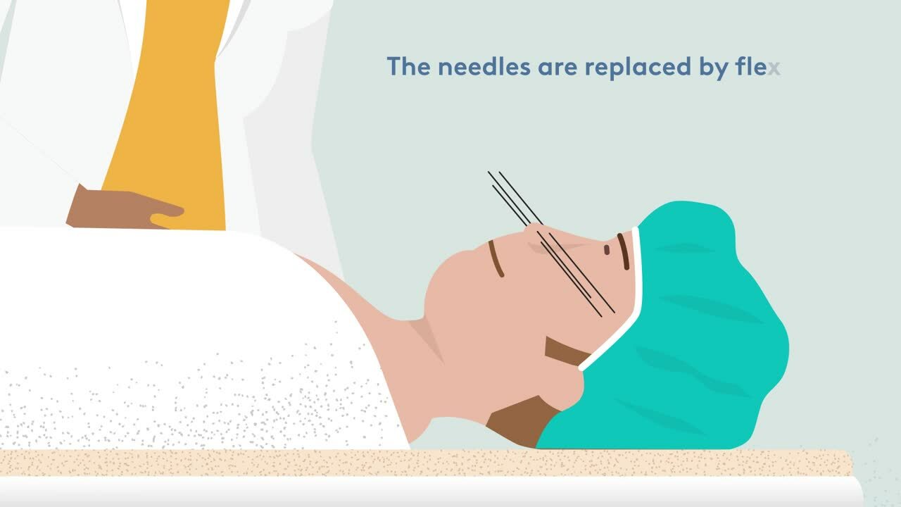
For lesions with depth greater than 5 mm and/or the tumor is in curved surfaces as in the face, an interstitial brachytherapy is indicated. The implantation procedure requires general or local anaesthesia. Often the flexible implant tubes are inserted and secured by fixation buttons or sutured to the skin.

Post-procedure monitoring
A follow-up appointment will be scheduled a few weeks after the last fraction has been delivered.
This appointment is to check that the treatment is going well and to monitor for any possible side effects. Typically follow up visits are scheduled every 3-6 months for the first year and thereafter once per year.9 This schedule of follow up does not depend on the type of therapy for skin cancer but is a general procedure when patients have had skin cancer. Also patients are recommended to regularly screen their skin for new lesions since the risk of developing new skin cancers is significantly higher after having had a first skin cancer lesion.

Brachytherapy:
The Precise Answer for Tackling Skin Cancer
References
- Guinot JL et al. Radiother Oncol 2018; 126:377-85. Available at: https://doi.org/10.1016/j.radonc.2018.01.013 Accessed June 2021
- Bussu F. et al. Brachytherapy 2021; 20(1):178-84. Available at: https://doi.org/10.1016/j.brachy.2020.08.008 Accessed June 2021
- Delishaj D et al. J Contemp Brachyther 2016; 8(6):533-40. Available at: https://doi.org/10.5114/jcb.2016.64112 Accessed June 2021
- Ouhib Z et al. Brachytherapy 2015; 14:840-58. Available at: https://doi.org/10.1016/j.brachy.2015.06.005 Accessed June 2021